scott@vtx-cpd.com
Forum Replies Created
-
AuthorPosts
-
Replying to Raquel M. 24/02/2025 - 16:21
No problem!
Let me know how you get on with the product!
Have a lovely weekend.
Scott 🙂
Hello everyone,
I went down a bit of a ketamine rabbit hole after you started this discussion—I didn’t realize people were using ketamine in such a sporadic subcutaneous way, and I think that’s really interesting.
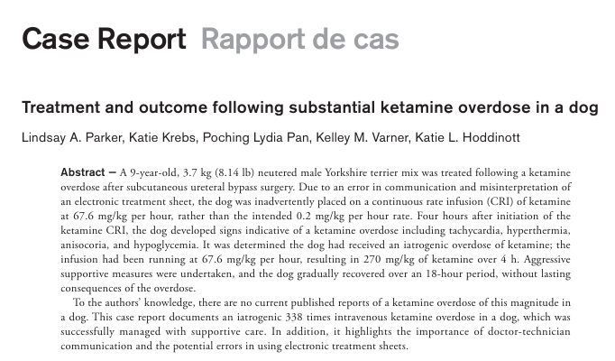
I also came across this case report that I thought was worth sharing. A dog accidentally received an extreme ketamine overdose—338 times the intended dose—due to a misinterpretation of an electronic treatment sheet. The dog was on a 67.6 mg/kg/hr CRI for four hours, receiving 270 mg/kg total, and developed tachycardia, hyperthermia, anisocoria, and hypoglycemia. Fortunately, with aggressive supportive care, he made a full recovery within 18 hours without lasting effects.
The case is published in Can Vet J. 2023 Mar;64(3):235–238 (PMC9979721, PMID: 36874544) and highlights both the potential resilience of patients in extreme circumstances and the importance of clear doctor-technician communication when using electronic treatment sheets.
Scott
Thanks again for the great question.
Are you using paracetamol a lot in your practice?
Scott 🙂
Replying to Raquel M. 25/02/2025 - 16:03
Hi Raquel,
Glad you found the information helpful!
Regarding the potential link between chronic paracetamol use and splenic tumours in dogs, I am not aware of any strong evidence supporting this association. While chronic paracetamol use in humans has been scrutinized for various long-term effects, including potential renal, cardiovascular, and hepatic impacts, there is no well-documented link to tumorigenesis in the spleen. In veterinary medicine, there is similarly no widely recognized data suggesting an increased risk of splenic neoplasia with chronic use.
If your colleague has seen a study or case series suggesting this link, I would be really interested to read more about it. Until there is more substantial evidence, I would consider this more of a theoretical concern rather than a proven risk?
Let me know if you come across anything on this; I’d be happy to discuss further.
Best,
Scott 🙂
Another brilliant video!
Scott 🙂
Replying to Josep B. 24/02/2025 - 10:11
Thank you!
Scott 🙂
Replying to Raquel M. 24/02/2025 - 16:45
Hi Raquel,
Thanks for your message, and I’m really glad the discussion has been helpful.
The 10–15 mg/kg dose every 8–12 hours, as recommended in Plumb’s, is generally considered safe and should provide effective analgesia in many cases. In otherwise healthy dogs, this dose is a reasonable starting point, particularly when used as part of a multimodal pain management approach. The Pardale-V dose used in the UK (33 mg/kg BID-TID) is higher but is based on a fixed paracetamol/codeine combination, which may alter its pharmacokinetics and clinical effect.
Efficacy at the lower dose depends on the type and severity of pain being treated. Paracetamol is a mild analgesic and antipyretic, and while it can be beneficial in musculoskeletal pain and chronic osteoarthritis, it may not be as effective for moderate to severe pain compared to NSAIDs, opioids, or adjunctive analgesics like gabapentin or amantadine. Some clinicians opt for the higher Pardale-V dose due to perceived improved efficacy, but there is a balance between achieving analgesia and minimizing potential hepatic or gastrointestinal side effects, particularly with long-term use.
If starting at 10–15 mg/kg and pain control seems inadequate, titrating within the safe range or adding adjunctive analgesia may be a better approach than immediately jumping to the higher dose. As always, patient monitoring, especially for any signs of toxicity, is key when using paracetamol in clinical practice.
Let me know if you’d like to discuss further.
Best,
Scott
Replying to Raquel M. 24/02/2025 - 16:55
Thanks for sharing your insights, Rachel! I completely agree, it’s so valuable to have evidence-based guidelines to refer to for these common but often frustrating cases.
Your approach to prazosin use aligns with a lot of the recent discussions I’ve seen. It’s interesting that you haven’t encountered rebound spasm, which makes me wonder if the weaning recommendation is more relevant for those using it frequently or at higher doses. I also appreciate your nuanced approach, reserving it for recurrent cases or those where preventing another obstruction is critical from a welfare or owner-retention perspective makes a lot of sense.
I really like that you incorporate the Ohio State Indoor Cat Initiative principles. It’s such a great resource for guiding owners through environmental modifications, which, as you pointed out, can be the hardest part when compliance is a challenge. Have you found any particular strategies from it that owners tend to be more receptive to?
Hydration strategies like Hydracare and pain management with gabapentin, buprenorphine, and maropitant are great adjuncts as well. I’ve seen Hydracare work well in some cases to lower USG, but uptake can be hit or miss depending on the cat. Do you find good owner compliance with it?
Along with the above guidelines, iCatCare has also released recommendations specifically for caregivers and owners, which might be a useful resource for client discussions: https://icatcare.org/resources/cat-carer-guide-urinary-tract-diseases.pdf
Really appreciate the discussion, these cases are always a puzzle!
Scott 😊
Replying to Josep B. 24/02/2025 - 10:08
It is a really interesting topic!
I am considering it more and more, I have been sing it with some chronic regurgitation/GI cases too.
Scott 🙂
Replying to Josep B. 24/02/2025 - 10:05
Thanks for sharing this, Josep—great summary of CK and its clinical relevance!
I’ve always been a bit unsure about how to use CK effectively, so it was really interesting to explore this further in your recent journal club. The Jones & Harcourt-Brown study was particularly insightful, showing how CK (alongside AST) can serve as a rapid screening tool for Neospora-associated meningoencephalitis. The high sensitivity and specificity at the 485 U/L cutoff make it a useful early marker, though I still wonder about its reliability in milder cases.
In practice, I find CK elevations are often non-specific, especially in medicine cases!
Thanks again.
Scott 🙂
Replying to Josep B. 24/02/2025 - 09:58
Really interesting.
Thank you for sharing.
Scott 🙂
Replying to Rosanna Vaughan 11/02/2025 - 12:36
Hello again.
I had a reply from my contact at Zoetis:
“The SPC’s for GB are on the VMD product database https://www.vmd.defra.gov.uk/ProductInformationDatabase/. Here you will find the most up to date SPC’s for any products registered in GB.”
I hope that helps!
Scott 🙂
Replying to Susana S. 25/02/2025 - 09:17
Thank you again Susana!
I hope you had a lovely holiday.
Scott 🙂
Josep, what are your thoughts on this paper?
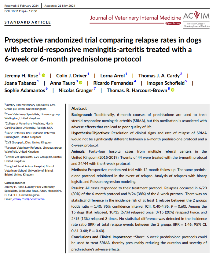
J Vet Intern Med. 2024 Jul-Aug;38(4):2221-2227 – a prospective randomized trial comparing relapse rates in dogs with SRMA treated with either a 6-week or 6-month prednisolone protocol.
A few key takeaways:
Both groups had similar response rates.
Relapse occurred in 30% (6-month protocol) vs. 38% (6-week protocol), but there was no statistically significant difference between them (P = 0.60).
Among relapsed cases, most dogs only relapsed once, with very few having multiple recurrences.
Suggests a shorter steroid course may be viable, potentially reducing adverse effects and improving quality of life.
Given the historical reliance on prolonged immunosuppression for SRMA, do you think this study is enough to shift treatment protocols towards shorter courses? Or are there limitations that make you hesitant to change standard practice?Does this change your tapering approach? What is your current approach to tapering in SRMA cases?
Scott 🙂
Replying to Josep B. 24/02/2025 - 10:13
“I know a really good paper on this topic… oh wait… did we write that?! 🙂
For anyone who somehow hasn’t read this masterpiece yet, let me summarize:
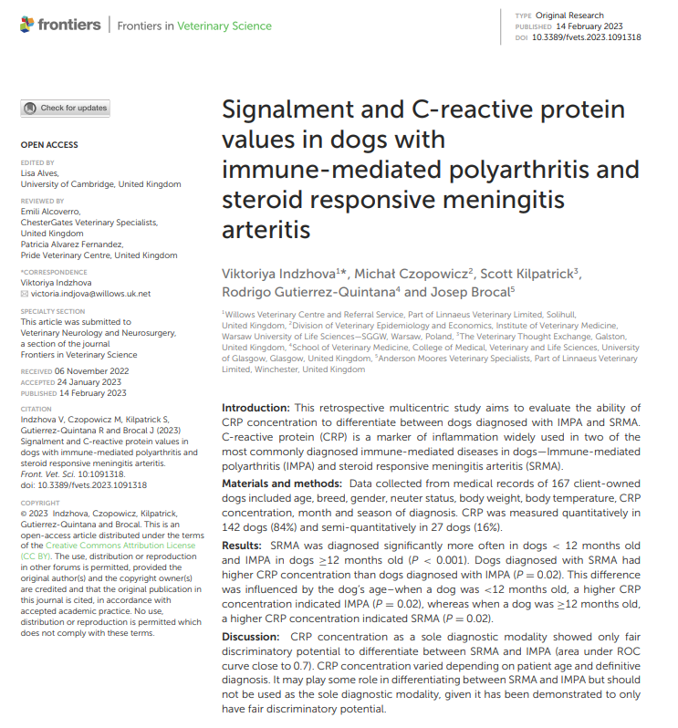
Front Vet Sci. 2023 Feb 14:10:1091318 – a fine example of scientific rigor, if I do say so myself! In this multicentric retrospective study, we analysed data from 167 dogs diagnosed with either SRMA or IMPA, looking at how CRP concentrations varied between the two diseases.
Key findings:
SRMA was significantly more common in dogs <12 months old, whereas IMPA was more frequent in dogs ≥12 months old (P < 0.001). Dogs with SRMA had higher CRP concentrations than those with IMPA (P = 0.02). However, CRP’s diagnostic role wasn’t as straightforward as expected—its predictive value actually shifted with age. In dogs <12 months old, higher CRP was paradoxically more suggestive of IMPA (P = 0.02). In dogs ≥12 months old, higher CRP was more indicative of SRMA (P = 0.02). When looking at overall discriminatory ability, CRP alone was only fair at differentiating between the two (ROC AUC ~0.7), meaning it shouldn't be used as a sole diagnostic tool. Take-home message: CRP is a useful marker of inflammation, but interpreting it in the context of SRMA vs. IMPA requires nuance—particularly considering patient age. It’s another reminder that clinical context always matters! Nothing like a bit of shameless self-promotion. Scott 🙂
-
AuthorPosts