scott@vtx-cpd.com
Forum Replies Created
-
AuthorPosts
-
Replying to Emma Holt 16/02/2023 - 12:40
Nailed the photo!
Have a great weekedn.
Scott 🙂
Replying to Emma Holt 16/02/2023 - 12:40
HAHAHAHAH! I knew you would!
I have even done helpful pictures!


Looking forward to seeing it!
Scott 🙂
Replying to Emma Holt 02/02/2023 - 13:48
Evening Emma!
In other news… you need to add a profile picture on here!
Peer pressure!
Scott 🙂
Replying to Kim Choo L. 08/02/2023 - 19:10
Hello Kim.
I hope you are well. Could you share the blood results? If you email them to me I can post them anonymously.
I think with such a low cobalamin I would be suspicious of significant small intestinal disease/chronic enteropathy.
It would be worth running a basal cortisol to rule out Addison’s. Have you tried a diet trial on this dog? If not it would be worth considering a hydrolysed diet. I would also consider Vivomixx as Jenny mentioned in her lecture.
The cobalamin will need longer if you are supplementing orally (12 weeks in some studies). The other option would be weekly injections. The cobalamin review is really helpful:
https://onlinelibrary.wiley.com/doi/full/10.1111/jvim.15638
The next steps, in this case, maybe endoscopy/biopsies. Steroids may be necessary. I would also consider the abdominal US.
The intermittent nature is not uncommon with some enteropathies. Will depend a bit on how much work the colon is doing with water reabsorption!
Hope that helps.
Scott 🙂
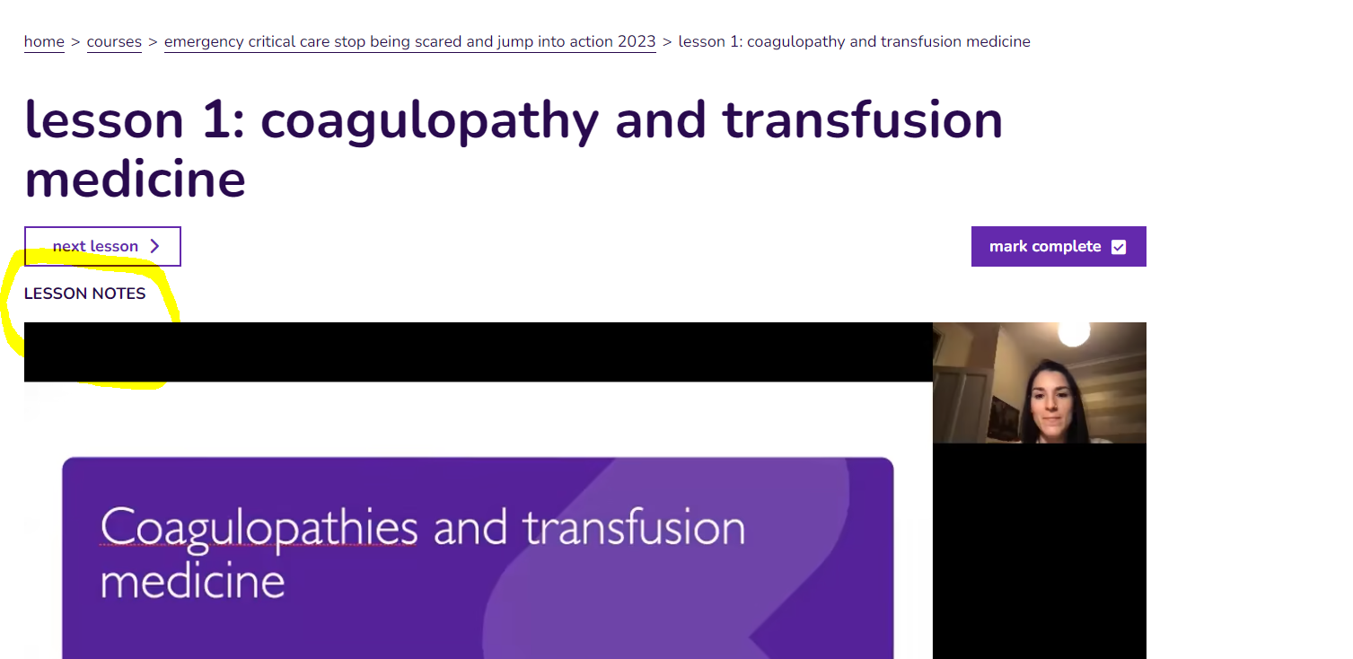
Replying to Alison S. 08/02/2023 - 17:12
Hey.
The notes are avaiable at the top left corner of the lesson. I have circled on the image.
Scott 🙂
Replying to Alison S. 08/02/2023 - 13:12
Hey Alison.
Let me know if you have any troubles finding the notes!
Scott 🙂
Replying to Liz Bode 02/02/2023 - 18:44
HAHAHAH!
Sorry, I should have read that properly!
Scott 🙂
Replying to Jennifer Cartwright 08/02/2023 - 23:04
Really interesting Jenny!
I look forward to seeing those results!
Scott 🙂
Hey Jenny.
Thank you for letting everyone know. Thank you so much for the brilliant course so far.
Sending lots of love.
Scott 🙂
Replying to Cristina M. 14/02/2023 - 00:00
Thank you Cristina!
You beat me to it!
Hope you are having a great week.
Scott 🙂
Replying to Francesca W. 10/01/2023 - 15:44
Hello Francesca.
I hope you are safe and well. I have no idea how I missed this question! I am so sorry for the delay!
I would normally use the 100mg dose as you mention. You could absolutely increase this dose if needed. The recommended dose is 5-10mg/kg PO q8-12 hours.
Interestingly, I was speaking to a colleague in the USA yesterday who works in a high volume spay and neuter clinic. They will use routinely ahead of elective procedures.
Hope that helps.
Scott
Replying to Sophie B. 07/02/2023 - 11:49
Sophie!
Lovely to hear from you. We seem to have a growing Aberdeen crew on the course.
I think CRI’s and acid-base fries the brains of the best of us, regardless of the time of day or night!
I really hope you enjoy the course.
Scott 🙂
Replying to Holly U. 06/02/2023 - 20:53
Hello Holly!
Welcome to the course. Thank you so much for learning with us!
I am pleased to hear that your heart lies in internal medicine… mine too!
I really hope you get lots out of the course. Let us know if you have any questions.
Scott 🙂
Hello Tessa.
Lovely to hear from you and great question!
For FNA’s I am mostly concerned about making sure they have enough platelets before sticking needles anywhere. There can be lots of reasons for platelets to be mildly decreased. As Jenny mentioned, with a mild/moderate decrease in platelets on the automated count but lots of platelets clumps, you would be reassured platelet number is probably OK. I would start to be concerned about the risk of bleeding when platelet number was below 50 10^9/L (without clumps) and really worried below 30 10^9/L.
Overall, I would not routinely run secondary coagulation parameters (PT and aPTT) before performing splenic (or other abdominal) FNA’s.
Hope that helps. Have a lovely week!
Scott 🙂
I agree with Emma!
Random question Liz… what do hyperdynamic pulses actually tell us?
I mostly think of them in IMHA cases!
Scott 🙂
-
AuthorPosts